Platelet-Rich Plasma Therapy
Platelet-rich plasma (or PRP) therapy is gaining more and more attention in both the regenerative medicine as well as mainstream medical fields now. It was first used in the 1970s with its use increasing through the 80s and 90s but common use in clinics only really started in the 2000s.
Blood is made of blood cells floating around within plasma. Plasma is the liquid part of blood separate to the cells which carries many nutrients, proteins, hormones, etc. Of the cells, 93% are red blood cells (RBCs), 6% platelets, 1% white blood cells (WBCs). Platelets are known to contain a multitude of growth factors contained within granules inside the cell as demonstrated below.
An electron microscopic image of an ultrathin (70 nm sample) PRP fraction. In the red circles, individual non-activated platelets are visible. In the blue circles, platelets have released their content following platelet activation. A single, non-activated, platelet at magnification ×7000 visualizes the three different platelet granules. Abbreviations: α: alpha granule; DG: dense granule; L: lysosome (Everts et al, 2023)
The normal function of a platelet is that when a platelet contacts any surface that is not the inside of a blood vessel it will trigger activation of the platelet (and release of granules) which will initiate blood coagulation (clotting) pathway as well as the release of growth factors. It is these growth factors that are potent recruiters of immune cells (mostly white blood cells) with the typical goal of protecting the body from potential invaders, as well as stimulating the healing cascade.
An illustration of an activated platelet, indicating pro- and anti-angiogenic platelet constituents released from α and dense granules. Abbreviations: TGF-β1: transforming growth factor beta 1; VEGF-A: vascular endothelial growth factor-ligand A; SDF-1α: stromal cell-derived factor-1 alpha; Ang-1: angiopoietin-1; IL-8: interleukin-8; MMP: matrix metalloproteases; PMP: platelet microparticles; TNF-α: tumor necrosis factor-alpha; 5-HT: serotonin; PDGF-BB: platelet-derived growth factor-BB; bFGF: basic fibroblast growth factor; TSP-1: thrombospondin 1; TIMP: tissue inhibitor of metalloproteinase; PF4: platelet factor 4; RANTES: regulated by T-cell activation and probably secreted by T cells (Everts et al 2023)
It is the latter effect that we are utilising to stimulate healing in a chronically injured tissue. However, it has been shown that injecting red blood cells into tissue can impair the healing response. With regards to leucocytes, some can have some detrimental effects on healing, particularly Neutrophils. However, other leucocytes, such as monocytes and lymphocytes, can potentially be beneficial in some scenarios.
What is involved?
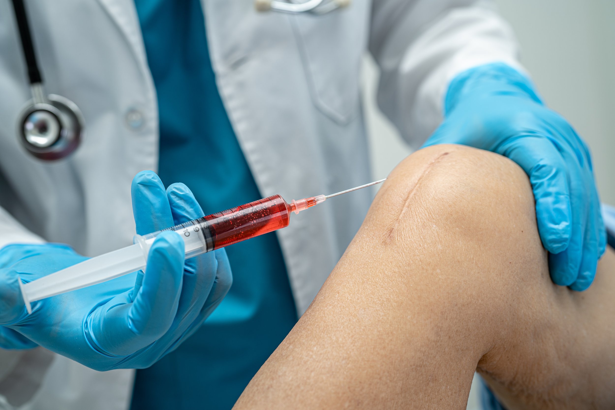
The process for PRP starts with a blood draw. This blood is collected into a sterile container and spun in a centrifuge. A 5–10-minute spin cycle applies a hugely exaggerated gravitational force on the blood and the blood contents that have the highest density (RBCs) sink to the bottom. The blood contents that have the lowest density (plasma) floats to the top. Blood contents with an in between density (platelets and leucocytes) will be in the middle. This separation by density allows us to collect the platelets +/- desired leucocytes with the plasma. To prepare PRP we need to separate off some of the plasma as mixing all the platelets with the reduced plasma volume is what makes it “platelet-rich”.
The exact concentration of platelets in the final injectate depends on a few factors. These include the patients baseline platelet count as well as the desired volume require to treat the injured tissue. Usually this will result in a concentration factor of between 3-9x the platelet concentration of the blood that has been drawn. However, factors like the patients baseline platelet count is important. Normal blood platelet levels at 150-450 x 109 per L. This means that if 2 people come in for a PRP treatment and patient 1 has a count of 150 and patient 2 has a count of 450, then both platelet counts are “normal”. However, since all PRP kits use a set volume of blood, this means patient 2 will get triple the platelet dose that patient 1 got. Therefore, the clinician should use a small volume of PRP for patient 1 so that the treated tissue gets an adequate concentration of platelets to actually stimulate adequate healing. In patient 2’s case, they may want to use a more generous volume to treat a larger area or maybe even avoid an excessive flare of pain during and after the injection. It is factors such as these that can significantly affect a patients experience and effect with PRP treatment to an injured tissue.
What is the evidence for PRP therapy?
One of the issues with early research with PRP therapy is that researchers really had to guess as to how best prepare the blood for injection. How much blood and how many platelets was required? How much centrifugation? Liquid or gel-based separation systems? How many treatments? Even individual practitioner methods for treating individual conditions could vary. This led to variable results. However, over time the data has become increasingly clear in the PRP space that it reliably can help with healing but making sure that the preparation is done correctly is also critical to success rates.
Here is a summary of data to date in a graphical form from the website https://regenexx.com/blog/my-2024-prp-rct-infographic/ (Centeno, 2024). This website does an annual review of the scientific literature for PRP treatments by conditions treated and graphs the results (good and bad). In the image below the blue circles showing studies with superior benefit of PRP, red circles showing studies where PRP was inferior to the comparator and orange being no significant difference. On the website clicking on each circle with take you to the link for the original scientific article, which I recommend people do if they would like to know more detail. As you can see the overall trend is definitely that of superiority over other treatments in most studies, however, not all of them. Certain conditions, like Achilles tendinopathy seem to have a lower success rate than others. Whereas some conditions like plantar fasciitis, epicondylitis (tennis or golfers elbow), osteoarthritis seem to have a very reliable benefit. Overall, in my experience the success rate of PRP treatment is in the 80-85% rate when it is done properly and for the right conditions.
Not all PRP is created equal
It is not uncommon for a patient to present with a condition that one would expect to respond well to PRP treatment for them to say that it didn’t work. It is true that some people do not seem to get the healing benefits one would expect from a PRP treatment, although in my experience this group is a small minority. This is because every person is slightly different in their response to a given treatment and for whatever reason, the PRP was not right for them. However, in my experience more often than not its because the PRP delivered previously is not adequate for the job and there are 3 major reasons for this.
Firstly, there are not enough platelets of concentration of platelets to provide effective results. Secondly, during the blood draw the platelets can become activated (rather than activating on injection into the tissue as is intended) meaning by the time they are injected they do not have the same healing effect. Thirdly, the quality of the PRP product can have a role in the quality of the sample.
At Nexus Pain Management we use a liquid based buffy coat PRP system. This means that when the PRP is prepared we can literally see the platelets entering the syringe for injection so we can be certain that we are delivering platelets that we are intending to. Other gel-based PRP systems have no visual cues at the time of preparation whether platelets are present or absent in the sample.
It is hard for the lay person to know what constitutes good quality of poor quality PRP. One simple starting question is - how much blood did they take for the injection? If it was only a small 10mL tube, it is likely that the platelet count may not have been high enough to have a reliable effect.
This study by Everts etc al (2023) demonstrates that beneficial healing was much more reliable when total platelet dose was > 3.5 x 10⁹ (3.5 billion).
Everts et al. (2023) Angiogenesis and Tissue Repair Depend on Platelet Dosing and Bioformulation Strategies Following Orthobiological Platelet-Rich Plasma Procedures: A Narrative Review. Biomedicines. 2023 Jul 6;11(7):1922
To break this down, a normal platelet count (where 95% of the healthy population lie) is 150-450 x 109/L. This equates to 150-450 x 106/mL. So, if someone has a 10mL blood sample taken and has a low but still “normal” platelet count of 150 x 106/mL x 10mL = 1500 x 106 platelets or 1.5 billion platelets. As you can see above the trials with platelet counts at the 1.5 billion mark did not produce significant outcomes. If a person’s platelet count is right on the upper limit of normal at 450 x 109/L (or 450 x 106/mL), then a 10mL blood draw will provide 4500 x 106 platelets, which will be just above this 3.5 billion threshold. Therefore, only those patients with platelet counts on the upper end of normal will start to get close to the platelet count required to give reliable benefit with a 10mL blood sample.
At Nexus pain our default blood volume for a PRP treatment is 30mL of blood. We also routinely get a pre-procedure full blood exam with platelet count prior to the PRP to ensure we know how many platelets we are starting with and can adjust the treatment to the desired effect. As per the above equations if 30mL of blood is taken, even those with low normal platelets counts of 150 x 106/mL x 30mL = 4500 x 106 or 4.5 billion platelets. For someone with a platelet count of 450 x 106/mL this would equate to 13.5 billion platelets! Plenty for the desired effect.
However, we also need to make sure the platelet concentration is high enough to have effects and target platelet concentration is 1-1.5 x 10⁹ platelets per mL (Advanced Regenerative Medicine Institute, 2023). If required we can and will increase the blood draw volume to 60 or even 120mL to make sure there are enough platelets for the desired effect.
So by knowing the patient’s baseline platelet count, the area that we want to treat and how much volume we will need, we can tailor your PRP to deliver the effect that you need and give you the best chance of a positive outcome.
Finally different companies’ preparation methods can affect the platelet and growth factor count. Here is an example of the differences in growth factor concentrations for different PRP products.
Kushida et al (2014) Platelet and growth factor concentrations in activated platelet-rich plasma: a comparison of seven commercial separation systems. J Artif Organs. 2014 Jun;17(2):186-92
At Nexus pain management we have trialled several commercially available systems and keep an active eye on ongoing research and new products to ensure our patients are getting a quality PRP system.
All treatmetns are also performed under ultrasound guidance, to make sure we are delivering the platelets exactly where we are intending to.
-
Advanced Regenerative Medicine Institute (13 Jul 2023) Choosing the Best PRP System for Your Practice - Matt Murphy, PhD [Video] https://www.youtube.com/watch?v=xLAa4FBuOF0
Centeno C (9 Jul 2024) My 2024 PRP RCT Infographic. Regenexx. https://regenexx.com/blog/my-2024-prp-rct-infographic/ [Accessed 6/3/2025]
Everts PA, Lana JF, Onishi K, Buford D, Peng J, Mahmood A, Fonseca LF, van Zundert A, Podesta L. Angiogenesis and Tissue Repair Depend on Platelet Dosing and Bioformulation Strategies Following Orthobiological Platelet-Rich Plasma Procedures: A Narrative Review. Biomedicines. 2023 Jul 6;11(7):1922. doi: 10.3390/biomedicines11071922. PMID: 37509560; PMCID: PMC10377284.
Kushida S, Kakudo N, Morimoto N, Hara T, Ogawa T, Mitsui T, Kusumoto K. Platelet and growth factor concentrations in activated platelet-rich plasma: a comparison of seven commercial separation systems. J Artif Organs. 2014 Jun;17(2):186-92. doi: 10.1007/s10047-014-0761-5. Epub 2014 Apr 20. PMID: 24748436.


